3D Topographical X-ray Machine
Author(s): ABDULWAHAB F. ALAHMARI
*Corresponding author: Alahmari AF, Department of Radiology, Al- Namas General Hospital, Ministry of Health, Al-Namas City, Saudi Arabia, Tel: +966562428716; E-mail: afaa99@hotmail.co.uk
Citation: AF (2024) 3D Topographical X-ray Machine. Clin Img and Med Case Rep Vol.1 No.2
Copyright: ©2024 Alahmari AF. This is an open-access article distributed under the terms of the Creative Commons Attribution License, which permits unrestricted use, distribution, and reproduction in any medium, provided the original author and source are credited.
Introduction: The development of an imaging modality takes time, effort, and energy to make a new machine. Lack of new imaging machines is prominent these days; therefore, in this paper, a proposal of a new imaging modality that will help in imaging patients in emergency settings.
Purpose: The aim of this paper is to propose a new imaging modality using images of the whole-body X-rays from 8 directions and combine these images to create a 3-dimensional topographical image that allows to see the whole-body from different directions with a low radiation dose. Scrolling and rotating the 3D image to see different views of the same body part in order to make sure if there is any pathology in the picture or not.
Material and Methods: The idea was studied to determine what design would serve a better purpose. Many designs were removed due to overlapping between the X-ray tubes and detectors. The best theoretical design in the arrangement of the X-ray tubes and detectors was tested by using a piece of paper and drawing the X-ray tubes and detectors then gather the two sides of the paper to make a gantry.
Results: The result will be a 3-dimensional X-ray image of the whole body that can be rotated to see different angles and views.
Conclusion: This new imaging modality can be used in emergency settings, forensic imaging, or for imaging the gastrointestinal tract with contrast media to allow better visualization from different angles.
Keywords
3 Dimension, X-ray, Topographical imaging, Bodygram, Trauma
Introduction
The development of an imaging modality takes time, effort, and energy to make a new machine. Lack of new imaging machines is prominent these days; therefore, in this paper, a proposal of a new imaging modality that will help in imaging patients in emergency settings. There is a machine called the Lodox-Statscan machine, which is useless because when a frecture is detected or suspected a patient to have a fracture then the patient has to go to do another X-ray both views. This machine neglects the role of two in radiology (1). Anyhow, this paper will propose a better idea of an X-ray machine that will eliminate the need for the traditional X-ray machine. This machine will depend on linear scanning of the entire body of the patient from head to toe with 8 views that will be combined to generate one 3D topographical X-ray image check bones that might have a fracture from the other 7 directions.
Literature Review
Someone named Mikael Haggström made a GIF image of a transition between two X-ray images (AP and lateral view) of the chest on Wikipedia, which he titled “projectional rendered CT scan” for some reason (2). However, the 2D X-ray images (i.e., 8 images) will be used to form a 3D model or generated surface of the human body (i.e., a topographical imaging, not a tomographical imaging). 3D geometry and reconstruction to make the images overlap each other as one image that can be seen from different surfaces. For example, make a transition GIF image of AP, RAO, right Lateral, RPO, PA, LPO, left Lateral, and LAO. This is how the 3D topographical X-ray machine images will look like. It will give a whole-body linear scan from 8 directions. There are machines like the Lodox-Statscan machine that scans in two directions, but the image will have an overlap of body parts. Therefore, the scan is usually done in one direction (i.e. AP view only), which neglects the rule of two in radiology (i.e., at least two X-ray views for the affected area) (1). In breast imaging, the mammogram is replaced with Digital Breast Tomosynthesis (DBT) imaging, which depends on many angles.
Material and Methods
The methodology of this paper starts with a new idea of an X-ray machine that can help take many views with less radiation dose compared to CT scanners. The machine is called a 3D Topographical X-ray machine, which helps to see the whole-body of the patient with 8 views to generate an image that can be rotated and manipulated to zoom in and out to see a certain part of the human body. The idea came to the author when he noticed the need of many patients to have different views, but they could not move at the same time due to a severe body injury. The idea came as imaging patients with 8 views in one exposure for the whole body, which will eliminate the need for a CT scan to check the orthopedic structures. The idea was studied to determine what design would serve a better purpose. Many designs were removed due to overlapping between the X-ray tubes and detectors. The best theoretical design in the arrangement of the X-ray tubes and detectors was tested by using a piece of paper and drawing the X-ray tubes and detectors. Then the paper was then gathered from both sides to make a gantry and each tube must face a specific detector. After changing places, the best arrangement to avoid X-ray overlap is to allow each tube to expose the body and be detected by its designated detector away from other X-ray sources and detectors. After that, a search of the literature was done to see if there was any machine with a similar design to the one proposed in this paper and the result was negative. Eventually, the design is presented in this paper and the process of this study is highlighted (Diagram1). The idea behind this machine is to create a 3D topographical image of the human body.
Results
3D topographical X-ray machine’s design
The design of the 3 D topographic machine i s similar to that of CT scanners in order to have X-ray tubes and detectors for imaging in all 8 directions. The X-ray tubes are fixed in their places, which requires l ess technology, like the slip ring and other technologies that are needed in moving CT machines. Also, this machine will eliminate the need for positioning the patients, which in many cases is difficult. Some parts overlap with other parts, like the lower limb lateral images, but this issue will be solved by having four oblique views of each limb. Some views, like the craniocaudal image of the skull and the planter- dorsal of the feet, are impossible views to acquire for three reasons: 1) the overlap of these images with the rest of the body; 2) the distance of X-ray tubes from the detectors in these parts is long; and 3) there are no detectors or X-ray tubes placed in those directions. The 8 images are posted on a 3D surface using geometry to allow seeing the body from different directions by moving the computer’s mouse. The huge field of view due to imaging the entire body needs to be worked on in order to achieve a high- quality image. Rotating and zooming the X-ray images to allow easy manipulation of the picture is essential. Theoretically speaking, this machine could be built, but some issues might be faced, like X-ray scatter from one of the X-ray tubes into another tube’s detector. But filters could be instilled and equipped with X-ray detectors. Linear imaging technology that is used in the Lodox- Statscan machines must be adopted and improved to reduce their size to fit 8 of them (i.e., 8 X-ray tubes and 8 detectors) in the 3D topographical X-ray machine.
The 8 X-ray tubes are placed in 8 directions, as seen in (Figure1). The generated 8 images, as seen in (Figure 2), are combined into one 3D topographic image of the whole-body. In order to image the whole body, all the X-ray tubes and detectors must cover the body from head to toe. The X-ray tubes and detectors are arranged in a fashion that prevents any overlap of images or X-ray sources see (Figure 3). The X-ray tubes are located in the middle of the X-ray machine see (Figure 3). The X-ray tubes generate a fan-shaped X-ray that is detected by a detector on the opposite side. The positions of the X-ray tubes and detectors are far from each other to allow imaging without any interference from other sources. The generated images in 3D will somehow look like the collected frames from Haggström’s GIF image see (Figure 4).
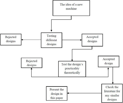
Diagram 1: A STARD chart to explain the methodology of making the design for this new idea of an X-ray machine.
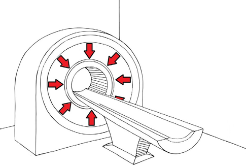
Figure 1: 3D Topographical X-ray tubes’ direction of acquisition of images.
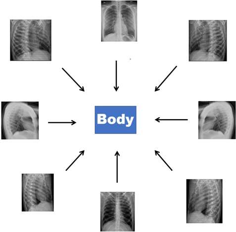
Figure 2: The d irection o f a cquiring t he X -ray i mages a nd what these images will show. After that, all these images will be combined into one 3D topographic image of the human's body.
Lodox-Statscan was developed from C-arm machines
This imaging modality is developed from the C-arm X-ray machine that is used in the theater room for surgery. The Lodox-Statscan machine allows orbital movement similar to the C-arm machine to allow different views, like oblique and lateral views (3). Both machines can’t have eight X-ray views combined into one 3D image.
Dual-Source CT
Some CT scanners for cardiac imaging have two sources of X-ray to image the heart very quickly, but they are huge machines due to the need to fit two moving X-ray sources in one machine. The issue in making the 3D topographical X-ray machine is fitting 8 X-ray tubes, 8 detectors, a massive cooling system, a data acquisition system, filters, collimators, a DC to AC converter, a high voltage multiplier, detector temperature control, etc. The machine gantry depth could be deep due to the need to arrange all the electrical parts inside the machine. The dual energy CT uses two layered detectors technology (i.e. top and bottom detectors). The low-energy photons are absorbed by a garnet scintillator based on yttrium in the upper layer, whereas high-energy photons are absorbed by gadolinium oxysulphide, which makes up the lower layer.
Discussion
There are many papers that are funded by the Lodox- Statscan company to publish good results and most of these papers do not include a conflict-of-interest declaration. However, there are negative results from their machine. For example, imaging of cervical spine injury detection rate is 57.1%, which is bad because the machine will provide an AP image of the cervical spine (4). The site of the fracture is associated with the detectability of the fracture on a Lodox-Statscan (5). The chest scan sensitivity was 44.4%, compared to a regular machine’s chest X-ray sensitivity, which is 72% (6,7). Lodox-Statscan company claims that the linear slot-scanning design produces a minimal distortion along the scan-axis and with foam placed on skeletons for anthropological research. This claim was found to have 85% agreement with dry bone measurements, which show that Lodox-Statscan is similar to a a regular X-ray machine’s (8). The distortion in the image increased when the foam was placed with skeletons in the Lodox-Statscan (8). The overall sensitivity of Lodox-Statscan for trauma imaging is 62%, compared to regular X-ray sensitivity for trauma, which is 75.4% (9,10). Additional radiographs were needed in 50% of the cases that were scanned by Lodox-Statscan (11). In one study where Lodox-Statscan was compared with X-ray, CT, and MRI, it was shown that Lodox-Statscan has a sensitivity of 5.3% for cervical spine trauma, which makes the machine insufficient for trauma imaging (12). In a study of 37 patients’ appendicular skeletal and pelvis trauma using CR, CT, and Lodox-Statscan, it was found that the Lodox-Statscan detected 46 abnormalities and missed 26 abnormalities that were detected on CR and CT (13). In a research paper that compared CR with Lodox-Statscan to measure the difference in the obtained information from Lodox-Statscan that did not appear on a CR machine. The result shows there is no difference in the obtained information (14). In maxillofacial trauma, the Lodox- Statscan was compared to a CT scanner, which showed that the Lodox-Statscan detected only 47 cases out of 245 patients, which represents 32% of the cases (15). Even if Lodox-Statscan was used to do a lateral view of the entire body, the image would be useless due to the overlap of the body’s parts over each other, which would make the image useless. The Lodox-Statscan was found to be insensitive in spine trauma imaging, which showed only 31 patients out of 332, which is 9% sensitivity only (16).
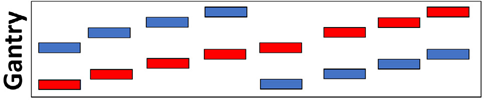
Figure 3: The arrangement of X-ray tubes (red) and X-ray detectors (blue) in this pattern to avoid overlapping of X-ray images. The acquisition process will be done in a helical fashion. Notice that the X-ray tubes are in the middle of the X-ray machine, where the detectors are on the periphery, to prevent X-rays from spreading outside the machine.
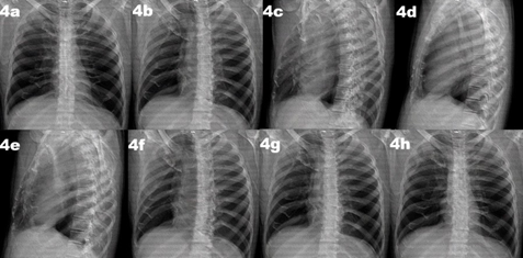
Figure 4: : Frames collected from the GIF image show different views that the 3D topographic imaging [3]. The software of the 3D topographical X-ray imaging will allow showing an image that can be rotated to see similar views, which are included here as frames from 4a to 4h.
In Dual-Source CT, the machine has a problem of fitting the parts in the gantry, which is described in many papers as “the detector’s size is limited by the gantry” (17). The rotation in Dual-Source CT takes 0.33 seconds, while the Lodox-Statscan takes 13 seconds to image the whole body (18,19).
Steps of reconstructing a 3D image from 2D images
- Calibration of the machine
- Collection of 8 X-rays by the data acquisition system
- Automatic background subtraction of the 8 views
- Build a 3D image on a geometric object
- Automatic reconstruction with little or no human intervention (iterative reconstruction) (20).
- Automatic correction of densities, image blur, silhouette, depth, and motion of the machine
- View the 3D image on the monitor.
Conclusion
The 3D Topographical X-ray machine is theoretically possible to be built, but with making the machine parts smaller than what we have in Lodox-Statscan. When this goal is achieved, this machine will be built to help emergency patients and even for other scans like GIT scans with contrast or forensic imaging.
Conflict of Interest
Authors declare no conflict of interest.
Funding
No funding.
References
- Abdulwahab Alahmari (2021) The rule of two in Radiology: An Update. O JRadio Med Img 4: 55-56.
- https://upload.wikimedia.org/wikipedia/commons/7/72/ Projectional_rendering_of_CT_scan_of_ thorax_%28thumbnail%29.gif.
- Bolton F (2017) Automated 3D reconstruction of Lodox Statscan images for forensic application.
- Chen RJ, Fu CY, Wu SC, Wang YC, Chung PK, Huang HC, et al. (2010) Diagnostic accuracy, biohazard safety, and cost effectiveness—the Lodox/Statscan provides a beneficial alternative for the primary evaluation of patients with multiple injuries. J Trauma Acute Care Surg 69: 826-830.
- Holdt FC, Pitcher RD (2019) An audit of the polytrauma fracture detection rate of clinicians evaluating lodox statscan bodygrams in two south African public sector trauma units. Injury. 50 :1511-1515.
- Mantokoudis G, Hegner S, Dubach P, Bonel HM, Senn P, Caversaccio MD, et al. (2013) How reliable and safe is full- body low-dose radiography (LODOX Statscan) in detecting foreign bodies ingested by adults? J Emerg Med 30: 559-564.
- Sargent W, Gibb I (2023) The sensitivity of chest X-ray (CXR) for the detection of significant thoracic injury in children exposed to blast. Injury 54: 1292-1296.
- Stull KE, L’abbé EN, Steiner S (2013) Measuring distortion of skeletal elements in Lodox Statscan-generated images. Clin Anat 26: 780-786.
- Deyle S, Wagner A, Benneker LM, Jeger V, Eggli S, Bonel HM, et al. (2009). Could full-body digital X-ray (LODOX- Statscan) screening in trauma challenge conventional radiography? J Trauma Acute Care Surg 66: 418-422.
- Dillon DG, Rodriguez RM (2021) Screening performance of the chest X-ray in adult blunt trauma evaluation: Is it effective and what does it miss? AJEM. 49:310-314.
- Deyle S, Brehmer T, Evangelopoulos DS, et al. (2010) Review of Lodox Statscan in the detection of peripheral skeletal fractures in multiple injury patients. Injury 41: 818-822.
- Rutsch N, Amrein P, Exadaktylos AK, Benneker LM, Schmaranzer F, Müller M, et al. (2023) Cervical spine trauma–Evaluating the diagnostic power of CT, MRI, X-Ray and LODOX. Injury. 54: 110771.
- Mulligan ME, Flye CW (2006) Initial experience with Lodox Statscan imaging system for detecting injuries of the pelvis and appendicular skeleton. Emergency radiology 13: 129- 133.
- Boffard KD, Goosen J, Plani F, Elias D, Herman P (2006) The use of low dosage X-ray (Lodox/Statscan) in major trauma: comparison between low dose X-ray and conventional X-ray techniques. J Trauma Acute Care Surg 60: 1175-1183.
- Deyle S, Evangelopoulos DS, Brehmer T, Zimmermann H, Exadaktylos AK (2011) Full body radiography (LODOX- Statscan) as a screening device for maxillofacial injuries, where CT scanning is not immediately available. Injury 42(1): 112-113.
- Häckel S, Hofmann E, Anwander H, Albers CE, Basedow J, Bigdon SF, et al. (2021) Anterior-posterior view by full- body digital X-ray to rule out severe spinal injuries in Polytraumatized patients. BMC emergency medicine 21: 1-10.
- Fletcher JG, Takahashi N, Hartman R, Guimaraes L, Huprich JE, Hough DM, et al. (2009) Dual-energy and dual-source CT: is there a role in the abdomen and pelvis? Radiol Clin North Am 47: 41-57.
- Flohr TG, McCollough CH, Bruder H, Petersilka M, Gruber K, Süβ C, et al. (2006) First performance evaluation of a dual- source CT (DSCT) system. European radiology 16: 256-268.
- Evangelopoulos DS, Deyle S, Zimmermann H, ExadaktylosAK (2011) Full-body radiography (LODOX Statscan) in trauma and emergency medicine: a report from the first European installation site. Trauma. 13: 5-15.
- Conference proceedings: Aharchi, M., Ait Kbir, M. A Review on 3D Reconstruction Techniques from 2D Images, editors. Springer, Cham. Innovations in Smart Cities Applications. Springer cham 2020.