Calcifications after Cerebral Infarction: Images in Clinical Radiology
Author(s): ABDULWAHAB F. ALAHMARI
Department of Radiology, Al-Namas General Hospital, Ministry of Health, Al-Namas City, Saudi Arabia
*Corresponding author: Alahmari AF, Department of Radiology, Al- Namas General Hospital, Ministry of Health, Al-Namas City, Saudi Arabia, Tel: +966562428716; E-mail: afaa99@hotmail.co.uk
Citation: Alahmari AF (2024) Calcifications after Cerebral Infarction: Images in Clinical Radiology. Clin Img and Med Case Rep Vol.1 No.2
Copyright: ©2024 Alahmari AF. This is an open-access article distributed under the terms of the Creative Commons Attribution License, which permits unrestricted use, distribution, and reproduction in any medium, provided the original author and source are credited.
Teaching Point: Calcifications in an area of the infarction after the event of cerebral infarction are a rare finding in stroke cases, and they might indicate a secondary hemorrhage, tumor, vascular malformation, or calcified granuloma.
Keywords: Calcification, Cerebral infarction, Computed tomography, Brain, Rare finding
Case History
A 55-year-old female patient with a history of a brain infarction before a year ago that caused a partial loss of sensation on the right side. She came to the hospital again, complaining of a chronic headache. A brain CT scan was performed and it revealed multiple calcifications in the infarction area in the left parietal lobe. The infarction is at the supra-ventricular level see (Figure 1) and at the level of the body of the lateral ventricles see (Figure 2). The calcification Hounsfield Unit (HU) measured 215.90 ± 19.50. The number of calcifications is 18 small pieces range in diameter from 0.05 mm to 3 mm. The infarction is located in the Middle Cerebral Artery’s (MCA) territory and in the watershed zone between the MCA and the Posterior Cerebral Artery’s (PCA) territory.
Comments
Calcification of cerebral infarction indicates a secondary hemorrhage, tumor, vascular malformation, or calcified granuloma [1]. A case series of three cases showed that calcifications formed in a timeframe of 4 months to 3.4 years post-infarction [1]. The calcium accumulation occurs due to the lack of plasma membrane integrity which keeps a low intercellular level against the high electrochemical gradient [1].
Conflicts of Interest
The author has no conflict of interests.
Abdulwahab F. Alahamri
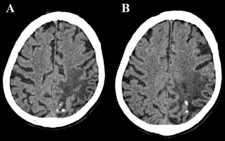
Figure 1: An axial CT scan of the brain at supraventricular level shows calcifications formed post-infarction
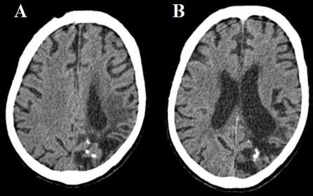
Figure 2: An axial CT scan of the brain at ventricular level shows calcifications formed post-infarction
Reference
- Kapila A (1984) Calcification in cerebral infarction. Radiology 153: 685-687.