Ophthalmology Findings on Brain Scans: A Pictorial Essay
Author(s): ABDULWAHAB F. ALAHMARI
Department of Radiology, Al-Namas General Hospital, Ministry of Health, Al-Namas City, Saudi Arabia
*Corresponding author: Alahmari AF, Department of Radiology, Al- Namas General Hospital, Ministry of Health, Al-Namas City, Saudi Arabia, Tel: +966562428716; E-mail: afaa99@hotmail.co.uk
Citation: Alahmari AF (2024) Ophthalmology Findings on Brain Scans: A Pictorial Essay. Clin Img and Med Case Rep Vol.1 No.2
Copyright: ©2024 Alahmari AF. This is an open-access article distributed under the terms of the Creative Commons Attribution License, which permits unrestricted use, distribution, and reproduction in any medium, provided the original author and source are credited.
The aim of this paper is to highlight some of the ophthalmological findings on brain scans. This paper will present uncommon findings that must be noticed during interpreting radiology scans. The eye balls or orbits generally, are in the blind spot during reading a brain scan. The prevalence of these findings is low among all patients, but still seen in a few cases
Keywords: Ophthalmology, Imaging, Brain, Scan, Blind spot
Pictorial Essay
This essay will highlight some ophthalmological findings on brain scans. Since the orbits are in the blind spot of brain scans, some ophthalmology findings could be missed. Here are some cases with low prevalence that could be missed or confused when interpreting brain scans.
Prosthetic Eye
Case presentation
This is a case of a 74-year-old female patient who came to the ER complaining of headaches and weakness of the left arm. A CT brain was requested to rule out stroke. The patient has an artificial eye on the left side as seen (Figure 1).
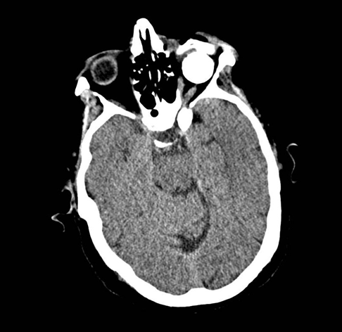
Figure 1: An Axial CT of the brain shows a hyper-dense round eye ball on the left side which indicates an artificial eye ball. Post hydroxyapatite sphere implantation after enucleation of the eyeball.
Prosthetic or artificial eyes are implanted in 0.1% of the population (1). In the UK, for example, 60,000 to 70,000 people have a prosthetic eye of all the population[2]. It appears as a round hyper-dense sphere. Sometimes it does not appear round, but a concave eye attached to the orbital muscles. Now, a new trend of 3D printers can make a prosthetic eye (1, 2). In some cases, the patient is blind in both eyes and they undergo a bilateral oculoplasty.
Phthisis Bulbi
Case presentation
This is a case of a 73-year-old male patient who came to the ER with dizziness and loss of balance. A brain MRI was requested to rule out any changes in the cerebellum. The left eye is affected with phthisis bulbi see (Figure 2).
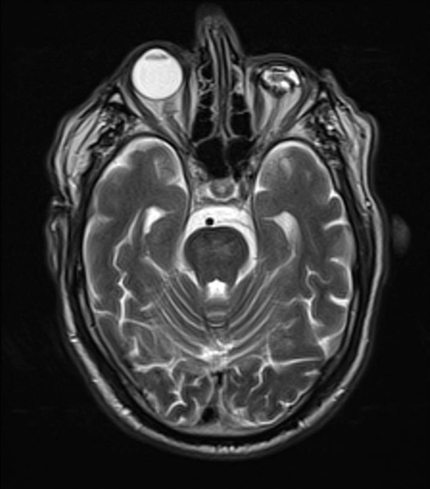
Figure 2: An axial MRI T2 sequence of the brain shows phthisis bulbi in the left eye ball.
Phthisis bulbi is the end stage or atrophied eye ball due to a severe insult. The prevalence of phthisis bulbi, for example, in one hospital in Nigeria is 0.45% out of 19,852 patients (3). The most common age group with phthisis bulbi is 65-85 years (4), meanwhile in younger ages, phthisis bulbi is rare and it happens due to malformations (4). The eye ball usually is reduced in size by 20 mm with posterior folded sclera. This intraocular fibrosis and shrinkage lead to an intraocular hypotension. Dystrophic calcification is seen in some cases which known as intraocular bones. Having calcification in the eye should not be confused with phthisis bulbi because calcification of the eye could happen for different reasons. The calcification could reach the globe, sclera, optic nerve, etc. The etiology of phthisis bulbi includes the following; infection, inflammation, trauma, retinoblastoma, chronic retinal detachment, persistent hyperplastic primary vitreous, etc.
Dislocated Lens
Case presentation
This is a case of an 86-year-old female patient who underwent a brain MRI to rule out any morphological changes in the pituitary gland. She has an old trauma which caused her a dislocated lens before 30 years ago and the patient did not want a surgery for her eye see (Figures 3-5).
The prevalence of the dislocated lens is 0.2% to 0.3% of the population (5). Dislocated lens or ectopia lentis occurs due to trauma, Weil-Marchesani syndrome, Ehlers-Danlos syndrome, homocystinuria, aniridia, syphilis, intraocular tumors, and Marfan syndrome.
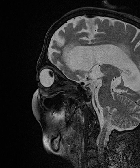
Figure 3: A sagittal MRI T2 sequence of the pituitary gland shows a dislocated lens on the left eye ball.
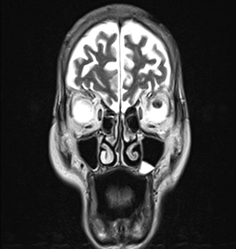
Figure 4: A coronal MRI T2 sequence shows a dislocated lens in the left eye ball.
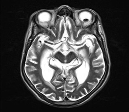
Figure 5: An Axial MRI T2 sequence of the pituitary gland shows a dislocated lens on the left eye ball.
Orbital Infection
Case presentation
This is a case of a 12-year-old female patient who underwent a root canal treatment and the treatment had some complications which resulted in cellulites on the left side. A CT scan was done to rule out any bone infiltration and extraocular muscles involvement which might be an orbital lymphoma or a mass which came back negative see (Figures 6-10).
Left eye proptosis due to post-septal orbital cellulitis (i.e. infection causing bulging eye). This type of infection usually treated with oral antibiotics. In this case, root canal re-treatment and antibiotics were recommended. Orbital infection count half of the primary orbital diseases? processes. Such infections can lead to proptosis, orbital apex syndrome, compressive optic neuropathy, orbital compartment syndrome, and irreversible visual loss. The patient usually presents with proptosis, chemosis, reduced visual acuity, and painful ophthalmoplegia. Orbital cellulitis happens as an extension of paranasal sinusitis. The orbital cellulitis appears on CT as a poor definition of orbital planes, soft tissue mass, edema, intraconal fat standing (inflammation), and abscess in advanced cases. When an abscess is present, a drainage of the content will be a necessity.
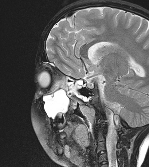
Figure 6: A sagittal MRI T2 sequence of the orbit shows the left orbital cellulites as a complication of sinusitis due to poor root canal treatment.
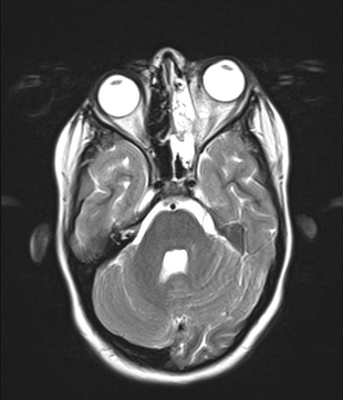
Figure 7: An axial MRI T2 sequence of the orbits shows a proptosis of the left eye ball due to the pressure made by the cellulitis in the left maxillary sinus.
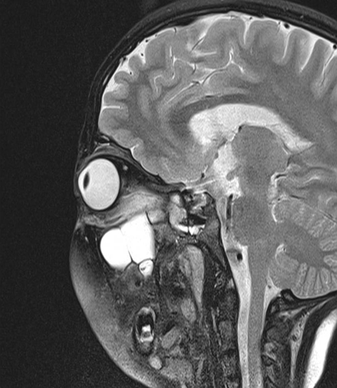
Figure 8: A sagittal MRI T2 sequences of the orbit shows the pressure made by the cellulitis in the left maxillary sinus.
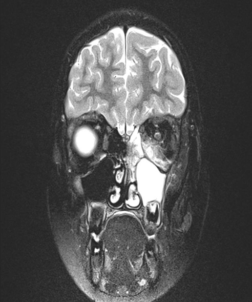
Figure 9: A coronal MRI T2 sequence shows the difference between eye balls level at the back of the right eye ball which indicates proptosis on the left.
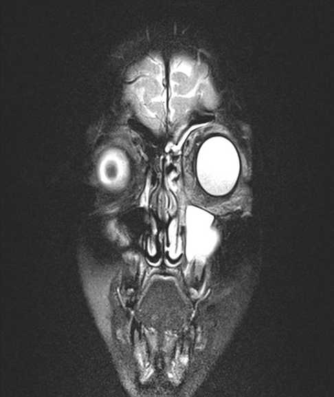
Figure 10: A coronal MRI T2 sequence shows the difference between eye balls level at the lens level which indicates proptosis.
Tortious Optic Nerves in Idiopathic Intracranial Hypertension (IIH)
Case presentation
This is a case of a 19-year-old female patient came to the ER with a severe headache and episodes of visual blurriness. A brain MRI was requested and it revealed bilateral optic nerves tortuousness due to an increased IIH see (Figures 11-13).
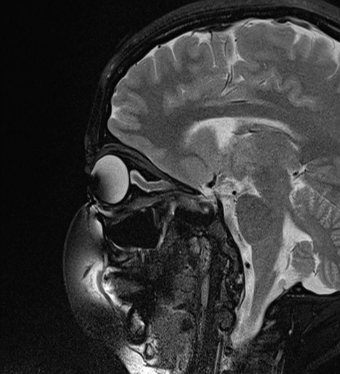
Figure 11: A sagittal MRI T2 sequence of the optic nerve shows a tortious optic nerve as an indication of Idiopathic Intracranial Hypertension.
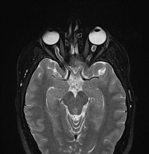
Figure 12: An axial MRI T2 sequence of the optic nerves show a tortious optic nerve which indicates an Idiopathic Intracranial Hypertension
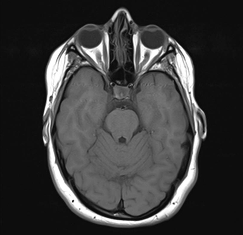
Figure 13: An axial MRI T1 sequence of the optic nerves show tortious optic nerves bilaterally which indicates an Idiopathic Intracranial Hypertension.
IIH is a syndrome where signs and symptoms of increased intracranial pressure is present with no mass or hydrocephalus. The IIH could result in a rapid visual loss, therefore, the term benign intracranial hypertension is frowned upon. Some cases of IIH occur due to venous stenosis, therefore, some authors call to use the term ?pseudotumor cerebri? which eliminates the idiopathic part, meanwhile others call to use the term Secondary Intracranial Hypertension (6).
Intraocular Lens Implants (pseudophakia)
Case presentation
A 76-year-old female patient complain of headaches and dizziness. A brains MRI shows a normal brain, but the left eye has an artificial lens implanted see (Figure 14).
A 30-year-long study showed that intraocular lens implants have a prevalence of 28.4 patient per million (7). Intraocular lens implants are safe under imaging using a 3T MRI machine (8). A study of 21 patients on a 7T MRI machine showed no heating effect or significant torque of the lens implants, but susceptibility artifact was seen in lenses that contain platinum (9). The artificial lens implants might be confused with cataracts, but clinically the patient had the surgery many years ago which rule out the cataract.
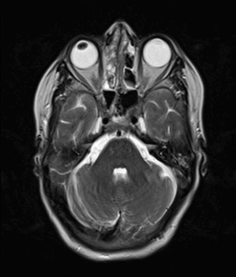
Figure 14: An axial T2-weighted MR image shows the lens on the left side is absent with a small isointense strip found on the right side as a postoperative change due to the artificial lens implantation
Ophthalmology Imaging
Ophthalmology practice uses more specialized in vivo noninvasive imaging techniques for assessment, monitoring, management and diagnosis. These techniques are: Optical Coherence Tomography (OCT) of the anterior and posterior segment, OCT angiography, corneal topography, confocal and specular microscopy, wide-field retinal imaging, and ocular ultrasonography which help in evaluating the orbital different morphology and function (10-21).
Conclusion
On brain scans, the orbits are in the blind spot and some findings in the eyes could be missed. These findings must be reported. Sometimes these findings are incidental and not the indication of the scan. Some of these findings are mostly common in older patients like; phthisis bulbi and prosthetic eye. The most common etiology for dislocated lens, phthisis bulbi, and prosthetic eye is trauma.
Reference
- Reinhard J, Urban P, Bell S, Carpenter D, Sagoo MS (2024) Automatic data-driven design and 3D printing of custom ocular prostheses. Nat Commun 15: 1360.
- Weaver T (2024) 3D-printed prosthetic eyes prove “life- changing” for patients. Engineering and Technology Magazine.
- Adewara BA, Badmus SA, Olugbade OT, Ezeanosike E, Adegbehingbe BO (2021) Distribution of phthisis bulbi and status of fellow eyes at a tertiary eye-care centre in Nigeria: a tenyear review. Afr Health Sci. 21: 437-444.
- Levin LA, Albert DM (2010) Ocular Disease: Mechanisms and Management: Expert Consult-Online and Print. Elsevier Health Sciences.
- Ascaso FJ, Huerva V, Grzybowski A (2015) Epidemiology, etiology, and prevention of late IOL-capsular bag complex dislocation: review of the literature. J Ophthalmol
- Degnan AJ, Levy LM (2011) Pseudotumor cerebri: brief review of clinical syndrome and imaging findings. Am J Neuroradiol 32: 1986-1993.
- Bothun ED, Cavalcante LCB, Hodge DO, Patel SV (2018) Population-based Incidence of Intraocular Lens Exchange in Olmsted County, Minnesota. Am J Ophthalmol 187: 80-86.
- Reiter MJ, Schwope RB, Kini JA, York GE, Suhr AW (2015) Postoperative imaging of the orbital contents. Radiographics 35: 221-234.
- Van Rijn GA, Mourik JE, Teeuwisse WM, Luyten GP, Webb AG (2012) Magnetic resonance compatibility of intraocular lenses measured at 7 Tesla. IOVS 53: 3449-3453.
- Fogel-Levin M, Sadda SR, Rosenfeld PJ, Waheed N, Querques G, Freund BK, et al. (2022) Advanced retinal imaging and applications for clinical practice: A consensus review. Surv Ophthalmol 67: 1373-1390.
- Romano V, Steger B, Ahmad M, Coco G, Pagano L, Ahmad S, et al. Imaging of vascular abnormalities in ocular surface disease. Surv Ophthalmol 67: 31-51.
- Hood DC, La Bruna S, Tsamis E, Thakoor KA, Rai A, Leshno A, et al. Detecting glaucoma with only OCT: Implications for the clinic, research, screening, and AI development. Prog Retin Eye Res 90: 101052.
- Romano V, Tey A, Hill NM, Ahmad S, Britten C, Batterbury M, et al. (2015) Influence of graft size on graft survival following Descemet stripping automated endothelial keratoplasty. BJO 99: 784-788.
- Borroni D, Romano V, Kaye SB, Somerville T, Napoli L, Fasolo A, et al. (2019) Metagenomics in ophthalmology: current findings and future prospectives. BMJ Open Ophthalmol 4: e000248.
- Liu S, Romano V, Steger B, Kaye SB, Hamill KJ, Willoughby CE (2018) Gene-based antiangiogenic applications for corneal neovascularization. Surv Ophthalmol 63: 193-213.
- Ren J, Gao X, Chen L, Lin H, Liu Y, Zhou Y, et al. (2022) Characteristics of the ciliary body in healthy Chinese subjects evaluated by radial and transverse imaging of ultrasound biometric microscopy. J Clin Med 11: 3696.
- Kim MS, Lim HB, Lee WH, Won YK, Nam KY, Kim JY (2021) Widefield swept-source OCT analysis of interocular symmetry of choroidal thickness in subjects with uncomplicated pachychoroid. J Clin Med 10: 4253.
- Zhang Z, Yao J, Chang S, Kanclerz P, Khoramnia R, Deng M, et al. (2022) Incidence and risk factors for Berger’s space development after uneventful cataract surgery: evidence from Alahmari AF. swept-source optical coherence tomography. J Clin Med 11: 3580.
- Parekh M, Leon P, Ruzza A, Borroni D, Ferrari S, Ponzin D, et al. (2018) Graft detachment and rebubbling rate in Descemet membrane endothelial keratoplasty. Surv Ophthalmol 63: 245-250.
- Romano V, Steger B, Myneni J, Batterbury M, Willoughby CE, Kaye SB (2017) Preparation of ultrathin grafts for Descemet-stripping endothelial keratoplasty with a single microkeratome pass. JCRS 43: 12-15.
- Ferrara M, Zheng Y, Romano V (2022) Imaging in Ophthalmology. J Clin Med 11: 5433.