Two Methods to Color Radiology Images of the Brain as an Example: Encoding Hounsfield Units with Hexadecimal Codes to Assign Colors for CT Scan or Using Prism in X-ray Cassatas or CT Detector to Convert the Light to the Real Colors in the X-ray or CT Images
Author(s): ABDULWAHAB F. ALAHMARI
*Corresponding author: Alahmari AF, Department of Radiology, Al- Namas General Hospital, Ministry of Health, Al-Namas City, Saudi Arabia, Tel: +966562428716; E-mail: afaa99@hotmail.co.uk
Citation: Alahmari AF (2024) Two Methods to Color Radiology Images of the Brain as an Example: Encoding Hounsfield Units with Hexadecimal Codes to Assign Colors for CT Scan or Using Prism in X-ray Cassatas or CT Detector to Convert the Light to the Real Colors in the X-ray or CT Images. Clin Img and Med Case Rep Vol.1 No.2
Copyright: ©2024 Alahmari AF. This is an open-access article distributed under the terms of the Creative Commons Attribution License, which permits unrestricted use, distribution, and reproduction in any medium, provided the original author and source are credited.
Introduction: All radiology images are gray scale images which does not show colors of the internal organs of the body. Seeing the real representation color of the images will help in diagnosis and prevent the use of autopsy in many cases.
Purpose: The aim of this paper is to assign a color for each Hounsfield unit in order to color CT scan images. The purpose to do such thing is to give real colors as much as possible for medical and forensic applications.
Methods: Two theoretical methods are discussed to see which one might be the better one to see real color representation of inner structure of the human body.
Results: The prism method might be better to have real colors coming out of the body and not assigned. The hexadecimal code needs collection of massive data to assign average degree of colors which is still not the real color.
Conclusion: The color of organs plays an important role in making the diagnosis of many conditions or to stop autopsy by seeing the organ color.
Keywords
Hexadecimal code; Color; Computed tomography; Hounsfield unit, Brain different by human compared to animals or to insects. But if everything is monochrome and we use the color-blind glasses, it will not show any colors. The eyes see things due to reflection of light off colors and objects to the human eyes based on their wavelength. The rod and three cons in the eye (i.e. Red, Blue, and Green (RBG)) collect these colors and show us mix of these colors. According to Johann Goethe hyperchromatism can lead to monochromatism (1). The depth of the image can be lighted by using darker colors which an old technique used since the renaissance era in arts. By knowing these principles that we can build on new techniques to color medical images in radiological machines in order to achieve seeing the colors of the inside the body without the need to do any autopsy. Having colors in radiology images will help in diagnosis, detection, and prognosis of different medical conditions.
This paper is a follow up of three published papers titled “Using CT scan to detect radiolucent foreign body (glass): An experimental study”, “Coloring CT scan images”, and “Propose image analysis tools to improve radiology interpretation” which are all written by the author of this paper. The ability to see colors of the inner organs in living patients could help as diagnostic tool if colors represent the real colors inside the body. Also, in post-mortem imaging it could help in knowing what went wrong to this deceased person by seeing the colors of organs. For example, some diseases could change the liver color to yellow instead of brownish red or other diseases that could change the color of the CSF to yellow from transparent like meningitis.
Coloring CT and X-ray as a start, is challenging task for many technical reasons. For example, how you can color transparent color like water on the computer, since there is no hexadecimal code for transparent color. In this paper, two theoretical methods will be proposed in hope this task will be achieved.
Methods
In this paper we propose two methods of coloring CT scan and X-ray images. The first option (which is the hardest), to color radiology images using hexadecimal codes for each part of the body. The second option (which is the less difficult option), to color radiology image using a prism and use the HU in CT or the density of the structure in X-ray image.
The first method will take coloring the brain task as a case study, but the prism needs an engineering and programming team effort to test this hypothesis. The theoretical ideas will be presented here since we lack the financial support.
In order to make the image looks realistic, some details and colors might be added to give a real look to the CT scan images. In forensic medicine, the colors of the organs indicate some pathologies by looking at the organ. By using a realistic coloring representation will eliminate the need for autopsy in few cases which the forensic pathologists needs to see signs for a disease that the pathologist suspects.
Results
The first method will not be accurate 100%, but the second method will rely on data coming from inside the body and gate these data into different channels. If coloring of the brain is done by assigning pseudo (fake) colors, then it will be useless since these pseudo colors will not represent what are the real colors inside the scanned subject’s brain. Follow of the blood inside the blood vessels of the brain will make the brain looks reddish due to the presence of blood. This issue can be solved by programming the scan by choosing if the person that will be scanned is deceased or still alive. If the person is deceased, then the computer should apply a blue (cold) image scale. This is done in order to reflect what the brain looks like inside the skull. If the person is still alive, then the computer must be using a red (hot) image scale to reflect how the patient’s brain looks like inside the skull. This representation is not accurate 100%, but it represents how the brain looks like from inside the skull to some extent.
In order to get the real colors exactly, it is very challenging task for many reasons. To do the coloring task, a CT scan must be done for a group of dead bodies who died by diseases that did not affect the brain then to do autopsy for those cadavers. The Hounsfield Unit (HU) measurement must be taken for every part of the brain like; brain surface, gray matter, white matter, meninges, and cerebrospinal fluid (CSF). The brains’ colors must be imaged with a camera to document the colors. Bear in mind, brains’ colors will change by death and by exposure to the air. Therefore, achieving the real colors will be impossible. What is possible is to try to make the most realistic colors possible to represents what is inside the skull of the scanned subject.
Programing the CT scan software must be made by the manufacturing companies of the CT scanners. This paper will help in directing what images should look like in most precise way possible. Pale colors could give a realistic picture of how organs look like inside a dead victim. There are view exception for this rule in many organs and many scenarios.
Another issue is that, there might be two organs and both have the same HU, but one looks brown and one looks pale red. This will be very difficult to differentiate between them by coloring them based on the HU only. As well, HU might not change in organs affected by a disease and remains the same as in a healthy organ. The color of the organ might change due to the disease, but the HU remains the same. Also, air and fluid have a different HU and both look dark on CT and both are transparent in real-life. Another example, is salt water and fresh water are different, but both have the same HU in drowning cases. Theoretically, in drowning cases, salt water will be thicker than salt water, but in reality, they can’t be differentiated on a CT scan of the chest. The blood is not differentiated from fluid in hemothorax and plural effusion based on density in the X-ray images, but they are differentiated by other radiological and clinical signs.
As well, glass inside the body is not easy to be detected by CT if it is a transplant glass. The glass which has low silica and lead does not appear on CT, while colored and thick glass with high silica and lead levels appears on CT images (2). Colors look-up table can’t help in detecting them (2). As well, colors look-up table is not useful to differentiate between healthy tissue (2). But colors look-up table could help in detecting abnormal tissues like in cases of prostate cancer (2). Another issue is that there are different diseases that change the organ color. For example, disease X causes the liver to look yellowish, disease Y causes the liver to look red, and disease Z causes the liver to look dark blue, but all of them have the same HU.
There are two papers proposed before to color CT images (3,4). The level of gray scale might be helpful to differentiate between structures. For example, air is very dark and fluid is less dark, so this level of difference on grey scale can be used to detect which is which?
Have you seen any cartoon show on TV? Usually when someone cry the tears will be colored white or aqua color. They color them white or aqua color because they can’t draw transparent color (i.e., the computer hexadecimal code of colors does not have the color transparent). According to Johann Goethe, white color is the darkest shade of transparent (1). He provides many evidences and observations in his book to support this claim. So white should be the closest to illustrate fluid on CT more than aqua color. But if the white color is used, the image will look like it has empty or no signal part. So, aqua which looks like water on cartoons is the best way to go.
Colors in human body change with change of normal physiology due to diseases. When blood stop circulating, it turns from dark red to dark blue (cyanotic). When the bile does not secrete due to an obstructed then the nails and eyes turn yellowish. So, change of the body’s colors indicates a pathology.
There is a possibility that every person has different shades of the same color of their inner organs. That could be similar to having anatomical variation, but in this case, it is inner organ color variations. In order to achieve real colors many cadavers need to be opened to take pictures then scan them then use the average between their colors to color on CT scan images. Some inner parts of the body might change when their bodies are opened and exposed to air.
There are two ways of coloring the human body on a CT scan. The first is assigning hexadecimal code and the second is using a prism. Let’s try both methods. In the first method we will pick a body part and try to assign hexadecimal code for normal appearance and pathological appearance.
A Case study of coloring the brain: assigning a hexadecimal code
When hexadecimal code is assigned to brain parts, the following table could be the closest to real representation of how the brain should looks like by using these hexadecimal codes see (Table 1 and Figure 1). Some of these colors might look a bit cartoonish if used without lighter and darker shades of the same color.
Table 1: Hexadecimal code assigned to brain structure to see how far brain colouring could be done.
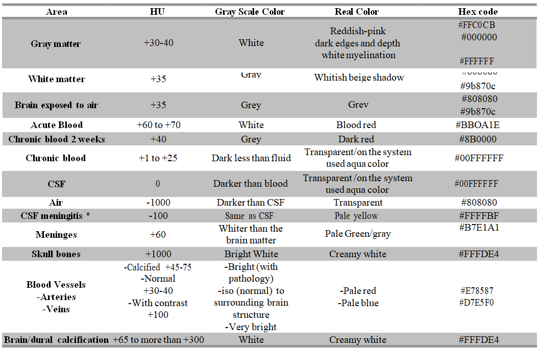
HU: Hounsfield Unit, Hex: Hexadecimal code, and CSF: cerebrospinal fluid. Note: Transparent color is not available on Hexadeci- mal Code (i.e. it can’t be done). * Meningitis cause leptomeningeal enhancement on CT, with normal CSF.
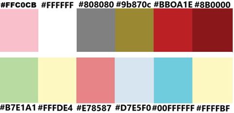
Figure 1: Reference for the colors in the hexadecimal code. Notice some of these colors do not represent the real color of inside the brain which will require to add depth to image by using darker colors as the old technique used since the renaissance era in arts.
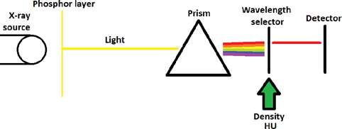
Figure 2: The prism method.
Here is the grade of colors that the hexadecimal codes above in the table shows. The issue that some brains might have different shades of the same color which is another obstacle. As presented above, the real colors can’t be achieved 100% accurately, but the closest coloring assigned, could be a good start.
Theoretical idea: The prism method
Another method is using the prism to convert the light generated from the phosphor layer in the cassette after absorbing the X-ray to convert it to the real colors see (Figure 2). This is a theoretical idea that can represent the real colors from the light to the cassette, but making the prism to fit into the cassette or CT detector will need good engineering. Prism can be made in small size then the wavelength of colors in the generated image can be assigned with HU, grey scale, or density of the X-ray to generate the real colors and the wavelength selector can block other colors from reaching the detector. Many small prisms are manufactured and spread on the detector or the receptor to convert all colors.
Discussion
Coloring CT scan image should not appear “cartoonic”, but rather imposed colors like a shadow. In a previous paper two methods were proposed based on two theoretical calculations, which goes as follows (3);
(R+B+G)/3= gray scale image (black)
Also, another equation state that;
((0.3 × R) + (0.59 × B) + (0.11 × G)) = gray scale image (brighter)
This method if reversed theoretically it will help in coloring images (4). These two equations when reversed then it can help in selecting the colors coming out of the prism. The issue this method depends on taking percents of red, blue, and green colors to color the image based on specific percentages as mentioned in the equation. But if the color of the prism was using these percentages to color the radiology image, it might show colors, but it needs to be tested. This means the colors of spectrum will take the three colors only and neglect the rest of the colors following the concepts that the eye needs only three colors to mix them to generate the rest of colors based on mixture of percentages of the main three colors (i.e. red, blue, and green). The percentages of coloring images will be adjusted according to the image input strength of these colors until the image colors reach a realistic level of colors or by using density or gray scale.
These methods can be incorporated then to test all of them to achieve coloring of radiology images.
Conclusion
These two methods are theoretical and both need further testing in order to achieve near real colors. Achieving the real colors inside the body without opening the body, it would be a difficult goal to achieve.
Conflict of Interest
Author declare no conflict of interest.
Funding
No funding
References
- Wolfgang von Goethe J (1810) Theory of Colours. Eastlake CL, trans. Mineola, NY: Dover Publications, 246.
- Alahmari A. (2020) Propose Image Analysis Tools to Improve Radiology Interpretation. IJR 7: 244-246.
- Alahmari A (2022) Coloring CT Scan Images. O J Radio Med Img. 5: 10-12.
- Alahmari AF (2023) Using CT scan to Detect Radiolucent Foreign Body (Glass): An Experimental Study. RRDI 2: 10- 58489.